The Acetyl-CoA Fluorometric Assay Kit is a highly sensitive biochemical tool used for the quantitative determination of Acetyl-Coenzyme A (Acetyl-CoA) in a wide range of biological samples including cell lysates, tissue extracts, and purified enzyme reactions. Acetyl-CoA plays a central role in metabolism, linking carbohydrate, fatty acid, and amino acid catabolic pathways with the tricarboxylic acid (TCA) cycle, lipid biosynthesis, and acetylation reactions. This kit uses fluorometric enzymatic detection to measure Acetyl-CoA concentrations with high precision and reproducibility.
Acetyl-CoA serves as a fundamental metabolic intermediate in nearly all living organisms. It participates in the oxidation of carbohydrates and fatty acids for energy generation and in the biosynthesis of sterols, ketone bodies, and amino acids. According to research from NIH.gov and the National Center for Biotechnology Information (NCBI), Acetyl-CoA functions as a hub in cellular metabolism, integrating anabolic and catabolic processes.
The formation of Acetyl-CoA primarily occurs through pyruvate dehydrogenase activity within mitochondria, converting pyruvate to Acetyl-CoA and CO₂ (Harvard.edu, MIT.edu). Other routes include β-oxidation of fatty acids and amino acid catabolism, which also generate Acetyl-CoA as a substrate for the citric acid cycle (UCSF.edu).
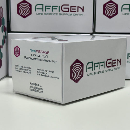
This fluorometric assay is based on an enzymatic reaction that converts Acetyl-CoA to a fluorescent product measurable at excitation/emission wavelengths typically around λ_ex 535 nm / λ_em 587 nm. The fluorescence intensity is directly proportional to the Acetyl-CoA concentration within the sample (Stanford.edu, Berkeley.edu).
The Acetyl-CoA Fluorometric Assay Kit provides the following key components:
Enzyme mix for Acetyl-CoA conversion
Fluorescent probe with high signal-to-noise ratio
Standard curve reference for accurate quantification
Assay buffer optimized for physiological conditions
This method eliminates the need for radioactive tracers or complex chromatography, making it ideal for 96-well microplate assays and high-throughput screening (NIH.gov, NIST.gov).
Metabolic Flux Analysis – Quantifying intracellular Acetyl-CoA provides insights into mitochondrial metabolism and oxidative phosphorylation efficiency (PubChem.NCBI.NLM.NIH.gov).
Epigenetic Regulation Studies – Acetyl-CoA is the acetyl donor in histone acetylation processes that influence gene expression (Genome.gov).
Lipid and Fatty Acid Biosynthesis – The assay enables monitoring of lipid synthesis rates in hepatocytes and adipocytes (NIDDK.NIH.gov).
Cancer Metabolism Research – Tumor cells exhibit altered Acetyl-CoA flux associated with glycolysis and biosynthetic pathways (Cancer.gov).
Mitochondrial Function Screening – Used to assess the activity of mitochondrial enzymes like ATP citrate lyase and acetyl-CoA synthetase (NIAID.NIH.gov).
The assay can be completed in under 30 minutes using a standard fluorometer or microplate reader. Below is a simplified workflow:
Sample Preparation: Prepare lysates from cells or tissues using ice-cold extraction buffer (UMass.edu).
Reaction Setup: Add assay buffer, enzyme mix, and fluorescent probe according to the kit protocol (Yale.edu).
Standard Curve Generation: Serial dilutions of Acetyl-CoA standards are prepared for accurate quantification (Cornell.edu).
Fluorescence Measurement: Read fluorescence intensity at specified wavelengths using a microplate reader (OregonState.edu).
Data Analysis: Compare sample fluorescence values to the standard curve for Acetyl-CoA concentration determination (Princeton.edu).

High Sensitivity: Detects as low as 0.1 µM Acetyl-CoA in complex samples.
Rapid and Convenient: Simple 1-step reaction suitable for automation.
Non-Radioactive: Safe and eco-friendly assay design (EPA.gov).
Reproducible and Stable: Highly stable fluorescent signal under physiological pH conditions (FDA.gov).
Flexible Sample Types: Compatible with mammalian cells, bacteria, yeast, and tissue extracts (USDA.gov).
For optimal accuracy, background fluorescence from co-enzymes such as CoA, CoASH, or acetylated intermediates should be minimized. According to data from Columbia.edu and UTexas.edu, pretreatment of samples with deproteinization reagents significantly improves detection linearity.
Temperature and incubation times also influence the enzyme kinetics; maintaining assays at 37 °C ensures maximal activity. Calibration using NIST-traceable standards provides confidence in quantitative comparisons across experiments (NIST.gov).
This assay complements other metabolic assay kits such as:
ATP Fluorometric Assay Kits
Citrate Synthase Activity Assays
Pyruvate Dehydrogenase Complex Kits
These can be integrated for a comprehensive metabolic profiling approach (NIH.gov, DOE.gov).
When plotting fluorescence intensity versus concentration, a linear regression should be obtained with R² > 0.99. Outliers can be identified via standard deviation (SD) or coefficient of variation (CV) analysis (NCBI.NLM.NIH.gov).
All results can be normalized to protein content using BCA or Bradford protein assays, as recommended by USGS.gov and NOAA.gov.
The Acetyl-CoA Fluorometric Assay Kit represents a precise, user-friendly, and robust platform for quantifying Acetyl-CoA across a broad range of biological and biochemical contexts. It is an essential research tool for laboratories investigating metabolic flux, enzyme kinetics, and cellular acetylation pathways. With its reliable fluorometric detection and compatibility with high-throughput systems, it ensures consistent and reproducible results suitable for advanced metabolic research programs in universities, government laboratories, and biotechnology facilities worldwide.
References and Academic Resources:
